- Conditions
- Tarsal coalition
Tarsal coalition
Growth of the tarsal bones / talocalcaneal coalition
Introduction
Tarsal coalition is diagnosed when two tarsal bones have (partially) fused into one larger piece of bone. A tarsal coalition may be congenital or develop over time. A tarsal coalition does not always have to lead to problems. When symptoms do occur, pain and stiffness in the mid-foot are the most prominent.
Tarsal coalition disorder occurs in about 1-2% of the people. In the congenital variant, symptoms usually develop during puberty. Symptoms that develop later in life are more likely the result of rheumatoid arthritis or may develop after a traumatic joint injury.
Description of condition
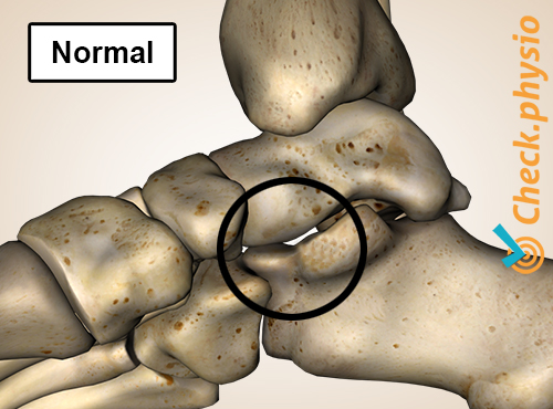
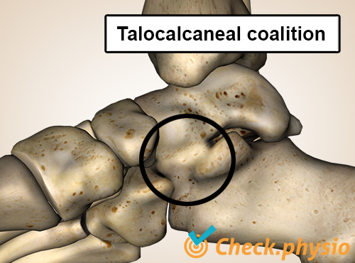
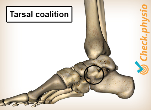
Normally, the tarsal bones in the foot consist of seven separate bones. In some people, two of these bones can fuse together to form a "coalition". The joint connection that is created may be made of bone, cartilage or connective tissue. If bone tissue, the connection will be very rigid. If connective tissue, the connection is still reasonably mobile. The stiffer the connection, the more likely it is to cause symptoms. Problems often arise elsewhere in the foot as well, because other joints are put under greater strain.
Tarsal coalitions are most frequently seen between the foot bones we refer to as the talus and calcaneus, and between the calcaneus and the navicular bone. A connection may also develop between other tarsal bones.
Cause and history
A tarsal coalition may be congenital but can also be the result of a joint disorder or an accident. When it’s a congenital disorder, it will be present in both feet in about 50% of the cases. The joint between the tarsal bones is already incorrectly formed at birth and develops into a tarsal coalition.
The moment at which the bones actually fuse depends on the bones involved. Not all bones fuse together equally fast. Most congenital tarsal coalitions are close to complete by the middle of puberty.
Symptoms primarily develop before the age of 20. That is once the bones have fused and the strain on the joints often becomes greater because of sports. For example, when someone just sprains an ankle and does not recover well, it may be because of the presence of a tarsal coalition.
As for non-congenital tarsal coalitions, this means that a disorder has seriously affected the joint. This might be rheumatism, a fracture or an infection. In a situation like these, the tarsal coalition can develop at any age.
Signs & symptoms
Possible symptoms of tarsal coalition are:
- Pain in the mid-foot.
- Stiffness of the mid-foot (stiff foot or feet).
- A flat foot that cannot be corrected.
- Frequent ankle sprains.-
Increased symptoms during or after activity.
Diagnosis
The correct diagnosis is sometimes missed if the patient is just asked some questions and given a physical examination. The physical examination can, however, reveal stiffness in certain joints. A flat foot may also be observed that cannot be corrected.
If the bones are completely fused together, the tarsal coalition can usually be seen clearly on an X-ray. A CT-scan or MRI can identify the problem in the first place when the connection between the bones does not (yet) consist of bone tissue.
Treatment and recovery
In order to reduce the symptoms caused by a tarsal coalition, non-operative remedies are applied first. Taking sufficient rest, fitting shoe inserts, taking painkillers and physiotherapy can all help.
If the above do not prove effective, surgery may be performed. During surgery, the two bones are separated again. The aim of this is to improve the mobility of the foot and to reduce the pain.
When a joint is seriously affected, or has fused over a large area, the joint may often be completely immobilized. This option can also be chosen if previous surgery did not help. The disadvantage is that the mobility of the foot will always be limited after this procedure. Activity patterns will have to be adjusted accordingly.
More info
You can check your symptoms using the online physiotherapy check or make an appointment with a physiotherapy practice in your locality.
References
Wiendels, D.R., Aarts, N.J.M., Steenmeijer, V.A. & Smeets, H.J. (2009) Herkenning van een tarsale coalitie. Klinische en radiologische aanknopingspunten Ned Tijdschr Geneeskd. 2009;153:A616.
Nugteren, K. van & Winkel, D. (2009) Onderzoek en behandeling van de voet Houten: Bohn Stafleu van Loghum.
Verhaar, J.A.N. & Linden, A.J. van der (2005) Orthopedie Houten: Bohn Stafleu van Loghum.
Brukner, P. & Khan, K. (2010) Clinical sports medicine McGraw-Hill: Australia. 3e druk.